Surgery Treatment

  • Home
  • Treatment
  • Surgery Treatment

ANATOMY OF NASAL SEPTUM

The nasal septum divides the nasal cavity into two halves.

Development of different parts of the ear

The nasal septum has three parts: -
Image

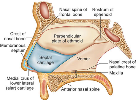
1. Columellar septum- It is formed of columella. It is made up of medial crura of alar cartilages joined together by fibrous tissue and covered on either side by skin.
a. Membranous septum- It consists of double layer of skin with no bony or cartilaginous support. It is situated between columella and caudal border of septal cartilage Both columellar and membranous parts are freely movable from side to side.
2. Septum proper- It consists of osteo-cartilaginous framework. It is covered by nasal mucous membrane.
Bony part: This is made up of:.

  • Postero-superiorly: The perpendicular plate of the ethmoid.
  • Postero-inferiorly: Vomer.

Cartilaginous part: It is formed by quadrilateral septal cartilage.

Articulation of the nasal septum:

The middle ear cavity and the auditory tube arise from first pharyngeal pouch called the tubotympanic sulcus, lined by endoderm.

    Superiorly:
    Sphenoid Rostrum.

    Anteriorly:
    Nasal bones, nasal spine of frontal bones.

    Inferiorly:

  • a) Nasal crest of the maxilla.
  • b) Nasal crest palatine process.
  • c) Anterior nasal spine of maxilla.
Arterial supply:

    Internal carotid system

  • 1. Anterior ethmoidal artery (branches of ophthalmic artery
  • 2. Posterior ethmoidal artery (branches of ophthalmic artery)

External carotid system:

  • a) Sphenopalatine artery (branch of maxillary artery) gives nasopalatine and posterior medial nasal branches.
  • b) Septal branch of greater palatine artery (branch of maxillary artery).

    Venous Drainage: :

  • a) Sphenopalatine vein.
  • b) Anterior and posterior ethmoidal veins.
  • c) Anterior facial vein

Nerve supply :

  • a) Trigeminal nerve including Anterior ethmoidal nerve.
  • b) Sphenopalatine ganglion.

Book Your Appointment